
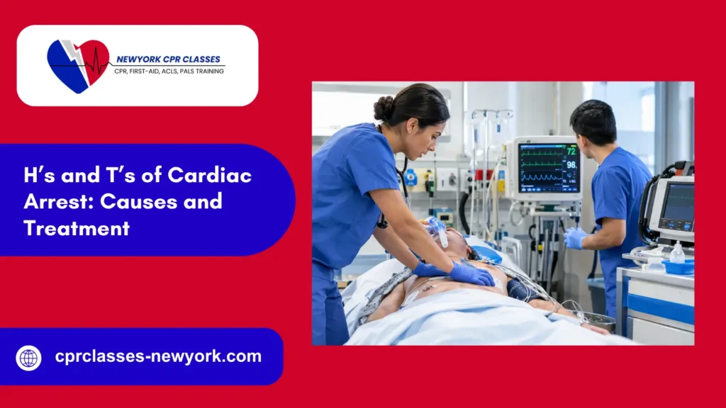
Cardiac arrest can occur due to several underlying problems, including low oxygen levels, severe blood loss, and blood clots. In Advanced Cardiovascular Life Support (ACLS), medical providers use the H’s and T’s mnemonic to recognize ten potentially reversible causes and guide clinical management during resuscitation.
This guide explains the five H’s and five T’s, their causes, and the treatment approaches used during cardiac arrest resuscitation.
What Are the H’s and T’s of Cardiac Arrest?
The H’s and T’s of cardiac arrest are a clinical memory aid used during Advanced Cardiovascular Life Support (ACLS) to help healthcare professionals assess why a patient’s heart has stopped pumping effectively. They provide a systematic way to consider underlying problems that may be treatable during resuscitation, particularly when the patient has pulseless electrical activity (PEA) or asystole.
Medical providers use this framework alongside cardiac rhythm assessment, high-quality CPR, and other indicated interventions to guide clinical decisions and address potential causes of cardiac arrest.
The Five H’s of Cardiac Arrest
| H | Reversible cause | What it means |
| 1. Hypovolemia | Low circulating blood volume | Severe blood or fluid loss reduces blood flow to vital organs. |
| 2. Hypoxia | Inadequate oxygen | Insufficient oxygen delivery can impair heart function and cause cardiac arrest. |
| 3. Hydrogen ion excess | Acidosis | Excessive acidity in the blood can impair normal cell function and the heart’s ability to work properly. |
| 4. Hypo- or hyperkalemia | Abnormal potassium levels | Potassium levels that are too low or too high can trigger dangerous heart rhythms. |
| 5. Hypothermia | Abnormally low body temperature | Severe cooling can slow heart activity and cause life-threatening arrhythmias. |
The Five T’s of Cardiac Arrest
| T | Reversible cause | What it means |
| 1. Toxins | Drug or chemical toxicity | Certain medications, drugs, or poisons can disrupt breathing, circulation, or cardiac function. |
| 2. Cardiac tamponade | Pressure around the heart | Fluid or blood surrounding the heart restricts its ability to fill and pump. |
| 3. Tension pneumothorax | Trapped air under pressure around a lung | Increasing pressure in the chest can reduce blood flow back to the heart. |
| 4. Pulmonary thrombosis | Blood clot in the pulmonary circulation | A pulmonary embolism can obstruct blood flow through the lungs. |
| 5. Coronary thrombosis | Blood clot in a coronary artery | An obstruction in a coronary artery can cause a heart attack and trigger cardiac arrest. |
The H’s and T’s are a clinical memory aid, not a replacement for the ACLS cardiac arrest algorithm. High-quality CPR, prompt defibrillation for shockable rhythms, and other indicated resuscitation measures must continue while the team evaluates and treats possible reversible causes.
The Five H’s of Cardiac Arrest Explained
1. Hypovolemia
Hypovolemia occurs when the volume of blood or fluid circulating in the body becomes too low. Severe blood loss, internal hemorrhage, major trauma, and significant dehydration can reduce the amount of blood returning to the heart. This limits cardiac output and may lead to circulatory collapse or cardiac arrest.
Signs and clues: A history of bleeding, trauma, vomiting, diarrhea, or severe fluid loss may suggest hypovolemia. Before cardiac arrest, a patient may have low blood pressure, a rapid heart rate, pale skin, or altered mental status.
Treatment considerations: Healthcare professionals address the source of blood or fluid loss and provide appropriate fluid resuscitation or blood products when indicated. Severe hemorrhage may require urgent bleeding control and transfusion.
2. Hypoxia
Hypoxia occurs when body tissues do not receive enough oxygen. It may result from airway obstruction, respiratory failure, drowning, severe lung disease, or other conditions that interfere with oxygen delivery. Prolonged oxygen deprivation can impair the heart and brain and lead to cardiac arrest.
Signs and clues: A history of breathing difficulty, choking, drowning, or respiratory disease can suggest hypoxia. Abnormal breathing, cyanosis, or low oxygen saturation before arrest may provide additional clues.
Treatment considerations: Healthcare professionals ensure adequate airway management, ventilation, and oxygen delivery. During cardiac arrest, they provide effective CPR and follow appropriate resuscitation protocols to support circulation and oxygenation.
3. Hydrogen Ion Excess (Acidosis)
Hydrogen ion excess means the blood has too many hydrogen ions, making it more acidic. Severe metabolic acidosis can occur with conditions such as shock, diabetic ketoacidosis, kidney failure, or prolonged inadequate tissue perfusion. Respiratory acidosis may develop when ventilation is insufficient to remove carbon dioxide.
Severe acidemia can interfere with cardiac contractility and the body’s response to resuscitation medications.
Signs and clues: The patient’s medical history, blood gas results, electrolyte measurements, and clinical circumstances may help identify acidosis. It can be difficult to distinguish acidosis caused by the arrest itself from an underlying condition that contributed to the arrest.
Treatment considerations: Management focuses on correcting the underlying cause and supporting effective ventilation and circulation. Sodium bicarbonate is not routinely administered for every cardiac arrest; its use depends on specific clinical indications and the applicable resuscitation protocol.
4. Hypokalemia or Hyperkalemia
Potassium helps control the electrical signals that regulate the heartbeat. Hypokalemia means the potassium level is too low, while hyperkalemia means it is too high. Either abnormality can disrupt cardiac electrical conduction and increase the risk of life-threatening arrhythmias.
Signs and clues: Kidney disease, dialysis, certain medications, gastrointestinal fluid losses, and known electrolyte abnormalities may raise suspicion. Electrocardiogram (ECG) changes and blood tests can help identify a potassium disorder.
Treatment considerations: Treatment depends on whether potassium is elevated or reduced. Severe hyperkalemia may require measures to stabilize cardiac cells and lower serum potassium, while hypokalemia requires appropriate potassium replacement. Management must account for the patient’s clinical condition and laboratory findings.
5. Hypothermia
Hypothermia occurs when the body’s core temperature falls below 35°C (95°F). Severe hypothermia can slow the heart rate, impair circulation, and increase the risk of dangerous cardiac rhythms. In some cases, it can lead to cardiac arrest.
Signs and clues: Cold exposure, immersion in cold water, prolonged exposure to low temperatures, and a low measured core temperature can suggest hypothermia.
Treatment considerations: Healthcare professionals provide appropriate rewarming, support oxygenation and circulation, and follow resuscitation protocols that account for severe hypothermia. Standard cardiac arrest decisions may need modification in profoundly hypothermic patients under specialist guidance.
The Five T’s of Cardiac Arrest Explained
1. Toxins
Toxins include drugs, medications, and chemicals that can disrupt breathing, blood pressure, or the heart’s electrical activity. An overdose or toxic exposure may cause respiratory depression, severe hypotension, dangerous arrhythmias, or cardiac arrest.
Signs and clues: Relevant history may include a suspected overdose, medication error, substance exposure, or access to toxic chemicals. Findings depend on the substance involved and may include abnormal pupils, altered consciousness, or characteristic ECG changes.
Treatment considerations: Management includes standard resuscitation, supportive care, and a specific antidote when one is indicated and available. The treatment approach depends on the substance, the patient’s condition, and toxicology guidance.
2. Cardiac Tamponade
Cardiac tamponade occurs when fluid or blood builds up in the sac around the heart, putting pressure on the heart and preventing its chambers from filling properly. This reduces the amount of blood pumped into circulation and can cause obstructive shock or cardiac arrest.
Signs and clues: Possible causes include chest trauma, bleeding around the heart, or medical conditions associated with pericardial effusion. Bedside cardiac ultrasound can help identify fluid around the heart and support the clinical assessment.
Treatment considerations: Emergency treatment may involve draining the fluid or blood compressing the heart through an appropriate procedure, such as pericardiocentesis or surgical drainage. The method depends on the cause and clinical circumstances.
3. Tension Pneumothorax
A tension pneumothorax develops when air accumulates under pressure in the space between the lung and chest wall. As air pressure builds up in the chest, it compresses the lung and makes it harder for blood to return to the heart, reducing blood flow throughout the body.
Signs and clues: Possible clues include chest trauma, sudden respiratory deterioration, reduced breath sounds on one side, and signs of obstructive shock. In an unstable patient, clinicians must identify the condition quickly and begin treatment without waiting for imaging tests.
Treatment considerations: Emergency decompression by a trained healthcare professional is required when tension pneumothorax is suspected in cardiac arrest. Definitive management commonly includes chest tube placement, according to the patient’s condition and local protocol.
4. Pulmonary Thrombosis
Pulmonary thrombosis in the H’s and T’s refers primarily to pulmonary embolism, in which a blood clot obstructs blood flow through the pulmonary arteries. A large pulmonary embolism can sharply increase pressure on the right side of the heart, reduce blood flow through the lungs, and cause cardiac arrest.
Signs and clues: A history of venous thromboembolism, recent surgery, prolonged immobility, active cancer, or other clotting risk factors may raise suspicion. Sudden shortness of breath, chest pain, or unexplained collapse before arrest may also provide clues.
Treatment considerations: Management depends on the clinical situation and available resources. In selected patients with cardiac arrest suspected to be caused by pulmonary embolism, systemic thrombolytic therapy may be considered. Other options, including catheter-based or surgical clot removal, may be appropriate in selected settings.
5. Coronary Thrombosis
Coronary thrombosis occurs when a blood clot obstructs a coronary artery, restricting blood flow to the heart muscle. This can cause an acute myocardial infarction, commonly called a heart attack, and trigger ventricular fibrillation, pulseless ventricular tachycardia, or other forms of cardiac arrest.
Signs and clues: Chest pain before collapse, a history of coronary artery disease, and relevant ECG findings may suggest an acute coronary event. The cause of cardiac arrest may remain unclear until medical providers can perform further tests and assessments.
Treatment considerations: Immediate cardiac arrest management remains the priority. Following return of spontaneous circulation (ROSC), urgent coronary evaluation and treatment, including percutaneous coronary intervention (PCI), may be required when indicated.
How Are the H’s and T’s Used During ACLS?
During Advanced Cardiovascular Life Support, healthcare professionals assess the patient’s cardiac rhythm, provide high-quality CPR, deliver defibrillation when indicated, and administer appropriate medications according to the resuscitation algorithm. At the same time, the team considers reversible causes that may be preventing the return of spontaneous circulation.
The H’s and T’s provide a systematic way to organize this assessment. For example, a patient with suspected severe blood loss may need urgent hemorrhage control and blood replacement, while a patient with suspected tension pneumothorax may require immediate chest decompression.
The team may review the patient’s medical history, examine the patient, and use ECG results, laboratory tests, or bedside ultrasound to help identify the cause of cardiac arrest when needed. These assessments should not unnecessarily interrupt chest compressions or delay defibrillation when it is needed.
Identifying a reversible cause is important, but not all causes can be confirmed immediately. Healthcare professionals must use clinical judgment, follow current resuscitation guidelines, and treat suspected conditions when the evidence supports intervention.
To apply the H’s and T’s effectively during cardiac arrest resuscitation, enroll in our American Heart Association (AHA) ACLS certification course at New York CPR Classes.
Why Are the H’s and T’s Important in Cardiac Arrest?
The H’s and T’s are important because some cardiac arrests involve conditions that require treatment beyond routine CPR and rhythm management. If a reversible cause remains untreated, the patient may not achieve ROSC or may experience another cardiac arrest after circulation returns.
Systematically considering these causes helps the resuscitation team:
- Identify potentially treatable conditions during cardiac arrest.
- Recognize problems that require specific interventions.
- Guide diagnostic assessment using available clinical evidence.
- Support decision-making during ACLS resuscitation.
- Address the underlying cause alongside standard cardiac arrest treatment.
The H’s and T’s are particularly useful in patients with pulseless electrical activity (PEA) and asystole, where identifying an underlying cause may be essential to management. However, medical providers should also consider these causes during other cardiac arrest rhythms when the patient’s condition suggests a possible reversible cause.
Strengthen Your ACLS Skills with New York CPR Classes
The H’s and T’s of cardiac arrest help medical providers identify potential causes of cardiac arrest that may be reversible with appropriate treatment. Alongside high-quality CPR and cardiac rhythm assessment, identifying and treating these causes is an important part of Advanced Cardiovascular Life Support (ACLS).
New York CPR Classes offers AHA ACLS training to help healthcare professionals develop their cardiac emergency response skills. Through this training, participants can practice rhythm recognition, follow resuscitation algorithms, and improve their clinical decision-making during cardiovascular emergencies. With multiple locations across New York, you can complete your ACLS training and get certified without unnecessary travel.
Frequently Asked Questions About the H’s and T’s of Cardiac Arrest
What are the 4 H’s and 4 T’s of cardiac arrest?
The 4 H’s and 4 T’s mnemonic lists eight possible causes of cardiac arrest that medical providers should consider during resuscitation. The standard ACLS mnemonic lists ten potentially reversible causes of cardiac arrest, divided into five H’s and five T’s, to help medical providers identify and treat them during resuscitation.
How can medical providers remember the H’s and T’s of cardiac arrest?
Medical providers can memorize the H’s and T’s by learning the five H causes and five T causes separately. Regularly reviewing the list and practicing ACLS scenarios can help them recall each cause and recognize the appropriate treatment during resuscitation.
What is the H’s and T’s mnemonic used for in ACLS?
The mnemonic helps medical providers identify potentially reversible causes of cardiac arrest and guide treatment during resuscitation. It is used alongside high-quality CPR, cardiac rhythm assessment, defibrillation when indicated, and other interventions in the ACLS algorithm.
What happens when someone flatlines and their heartbeat returns?
Flatlining is usually referred to as asystole, a cardiac arrest rhythm in which the monitor shows no detectable ventricular electrical activity. If circulation returns after resuscitation, the patient has achieved return of spontaneous circulation (ROSC). However, asystole is not a shockable rhythm, and medical providers must confirm the rhythm and follow the appropriate resuscitation algorithm.
Which H’s and T’s Can Be Identified Quickly After 20 Minutes of Resuscitation?
Hypoxia, hypovolemia, tension pneumothorax, and cardiac tamponade may be suspected through clinical assessment and bedside ultrasound when available. Electrolyte abnormalities may require blood tests for confirmation. The time spent performing CPR alone does not determine when resuscitation should stop.
Which H’s and T’s Can Be Identified Quickly During Cardiac Arrest Resuscitation?
Some causes of cardiac arrest can be identified through a quick patient assessment. Medical providers can check body temperature for hypothermia, look for signs of blood loss that suggest hypovolemia, and use bedside ultrasound to assess for tension pneumothorax or cardiac tamponade. Blood tests can confirm abnormal potassium levels, while the patient’s history and clinical assessment can help providers identify signs of low oxygen.
Is Trauma Included in the H’s and T’s of Cardiac Arrest?
Yes. Trauma is included as the 6th “T” in some expanded cardiac arrest frameworks, particularly when addressing cardiac arrest caused by severe physical injury. However, the standard adult ACLS framework focuses on five H’s and five T’s.
Which One of the H’s and T’s Represents a Potentially Reversible Cause of Cardiac Arrest?
Hypovolemia is one of the H’s and T’s that represents a potentially reversible cause of cardiac arrest. It refers to a severe loss of blood or body fluids that reduces blood circulation and oxygen delivery to vital organs.